Home
/ Stratified Columnar Epithelium Drawing - Histology Image Membranous Epithelium - In pseudostratified epithelium, nuclei of neighboring cells appear at different levels rather than clustered in the basal end.
Stratified Columnar Epithelium Drawing - Histology Image Membranous Epithelium - In pseudostratified epithelium, nuclei of neighboring cells appear at different levels rather than clustered in the basal end.
Stratified Columnar Epithelium Drawing - Histology Image Membranous Epithelium - In pseudostratified epithelium, nuclei of neighboring cells appear at different levels rather than clustered in the basal end.. 1 loose (areolar) ct a b 2. Columnar epithelium with goblet cells tissue / organ: Pseudostratified and transitional epithelia these two types of epithelia are difficult to classify using the shape of the cells in the surface layer and the number of. For example, by 8 weeks, it covers the lining of the stomach. The stratified columnar epithelium has multiple layers of cells in which the apical layer is made up of columnar cells while the deeper layer can be either cuboidal or columnar.
Epithelium covers the inner surface of the digestive tract. N, nucleus of columnar cell; Stratified columnar epithelium of mucous gland duct of the tongue; Stratified cuboidal epithelium organ choices: The stratified columnar epithelium has multiple layers of cells in which the apical layer is made up of columnar cells while the deeper layer can be either cuboidal or columnar.
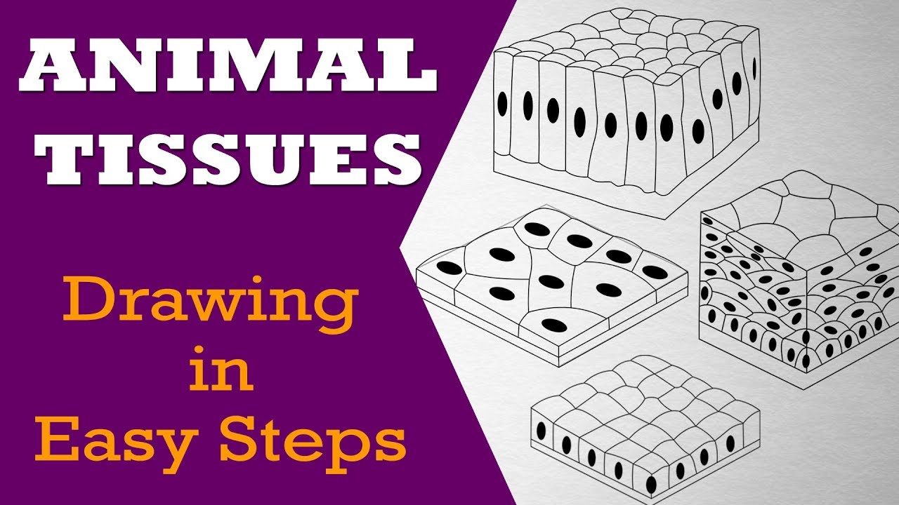
How To Draw Animal Tissues In Easy Steps 9th Biology Cbse Ncert Class 10 Science Youtube from i.ytimg.com It consists of several layers of cells with only the superficial layer having tall columnar cells. In pseudostratified epithelium, nuclei of neighboring cells appear at different levels rather than clustered in the basal end. Stratified cuboidal epithelium organ choices: Draw the stratified columnar epithelium seen in the largest draw the stratified columnar epithelium seen in the largest ducts and label your drawing. Fill in the 2 lines beneath the graphic. (points will be removed for graphics that do not point out and. Caitlin kenney stratified columnar epithelium may be found in the pharynx. In the intestines, it stays columnar but acquires microvilli to increase the surface area for absorption.
Stratified columnar epithelium of mucous gland duct of the tongue;
In the intestines, it stays columnar but acquires microvilli to increase the surface area for absorption. Epithelium covers the inner surface of the digestive tract. How many layers of cells are there? In this image, you will find basement membrane, stratified columnar epithelium, underlying connective tissue, urethra in stratified columnar epithelium anatomy. As in the case of other stratified epithelium, the cells in the deeper layers might be different than the layer on the top. If there is more than 1 layer; A pseudostratified epithelium is a type of epithelium that, though comprising only a single layer of cells, has its cell nuclei positioned in a manner suggestive of stratified epithelia.as it rarely occurs as squamous or cuboidal epithelia, it is usually considered synonymous with the term pseudostratified columnar epithelium. Pseudostratified and transitional epithelia these two types of epithelia are difficult to classify using the shape of the cells in the surface layer and the number of. Very abraded areas (mouth, anus, surface of skin) squamous tissue = squashed drawing simple vs. Stratified cuboidal epithelium organ choices: It consists of superimposed fusiform cells and is found in some parts of the male urethra. 1 loose (areolar) ct a b 2. The apical layer is columnar and the basal layer is cuboidal.
In this image, you will find basement membrane, stratified columnar epithelium, underlying connective tissue, urethra in stratified columnar epithelium anatomy. Several cell layers, basal cells usually cuboidal; Epithelium covers the inner surface of the digestive tract. In pseudostratified epithelium, nuclei of neighboring cells appear at different levels rather than clustered in the basal end. Stratified columnar epithelium of mucous gland duct of the tongue;
Table Of Tissues Epithelial Tissue Part 1 Design And Fill In A Table Of The Tissues Of The Human Body Include The Name Of The Tissue Type A Drawing Ppt Download from images.slideplayer.com The other layers may contain cells that are cuboidal and/or columnar, but the classification of the epithelium is based only on the shape of the outermost layer of cells. The drawings of histology images were originally. It starts as stratified squamous epithelium in the esophagus and changes to simple columnar epithelium in the stomach. N, nucleus of columnar cell; Organ function tissue function h. If there is more than 1 layer; As in the case of other stratified epithelium, the cells in the deeper layers might be different than the layer on the top. All the cells are attached to the underlying basement membrane, but the nuclei are at different heights, giving the appearance of a 'stratified' epithelium.
Pseudostratified columnar epithelium is a type of epithelium that appears to be stratified but instead consists of a single layer of irregularly shaped and differently sized columnar cells.
Several cell layers, basal cells usually cuboidal; N, nucleus of columnar cell; It starts as stratified squamous epithelium in the esophagus and changes to simple columnar epithelium in the stomach. Is each layer the same shape? Stratified squamous epithelia consist of multiple layers of cells with the outer most layer being squamous. Submandibular glands, conjunctiva, pharynx, anus, uterus, urethra, vas deferens organ draw an example. Pseudostratified ciliated columnar epithelium containing goblet cells lines most of the major airways. For example, by 8 weeks, it covers the lining of the stomach. All the cells are attached to the underlying basement membrane, but the nuclei are at different heights, giving the appearance of a 'stratified' epithelium. This is an example of pseudostratified epithelium, from the trachea. Organ function tissue function h. Superficial cells elongated and columnar. Draw a line showing where the basement membrane begins.
Pseudostratified columnar epithelium is a type of epithelium that appears to be stratified but instead consists of a single layer of irregularly shaped and differently sized columnar cells. (points will be removed for graphics that do not point out and. It consists of several layers of cells with only the superficial layer having tall columnar cells. For example, by 8 weeks, it covers the lining of the stomach. 1 loose (areolar) ct a b 2.
File Anatomy And Physiology Of Animals Stratified Squamous Epithelium Jpg Wikimedia Commons from upload.wikimedia.org Pseudostratified columnar epithelium is a type of epithelium that appears to be stratified but instead consists of a single layer of irregularly shaped and differently sized columnar cells. Stratified columnar epithelium is a type of epithelial tissue composed of two or more layers of columnar epithelial cells.epithelial tissue is one of the body's primary tissues and lines the surfaces and insides of structures in many parts of the body. This is an example of pseudostratified epithelium, from the trachea. The apical layer is columnar and the basal layer is cuboidal. For example, by 8 weeks, it covers the lining of the stomach. As in the case of other stratified epithelium, the cells in the deeper layers might be different than the layer on the top. Also draw some of the tissue (probably connective tissue) below the epithelial layer. Sweat glands organ draw an example.
N, nucleus of columnar cell;
Caitlin kenney stratified columnar epithelium may be found in the pharynx. Is each layer the same shape? All the cells are attached to the underlying basement membrane, but the nuclei are at different heights, giving the appearance of a 'stratified' epithelium. Ducts and label your drawing. Draw the stratified columnar epithelium seen in the largest draw the stratified columnar epithelium seen in the largest ducts and label your drawing. Look at the nuclei of the epithelial cells and notice that there are several layers of them. Columnar epithelium with goblet cells tissue / organ: Stratified cuboidal epithelium organ choices: Epithelium covers the inner surface of the digestive tract. Submandibular glands, conjunctiva, pharynx, anus, uterus, urethra, vas deferens organ draw an example. Look at and draw tissues on page 66. Stratified columnar epithelium organ choices: Several cell layers, basal cells usually cuboidal;

